



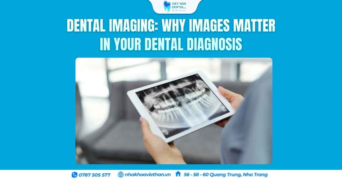
A dental examination involves more than simply looking at your teeth. Some oral health problems can be detected visually, while others may be hidden beneath the gums, between teeth or inside the jawbone.
Dental imaging helps dentists gather additional information, explain findings clearly and develop a treatment plan suited to each patient.
At Viet Han Dental Clinic in Nha Trang, diagnostic technologies are used when clinically appropriate to support accurate assessment and personalised dental care.
Nội dung
- What Is Dental Imaging?
- 1. Detecting Problems That Are Difficult to See
- 2. Supporting More Precise Treatment Planning
- 3. Helping Patients Understand Their Dental Condition
- 4. Monitoring Treatment Progress
- 5. Improving Communication Between Dental Professionals
- Is Dental Imaging Safe?
- Dental Imaging at Viet Han Dental Clinic
- Understanding Your Smile Starts With the Right Diagnosis
What Is Dental Imaging?
Dental imaging refers to the use of photographs, X-rays and digital scans to record and evaluate the teeth, gums, bite and surrounding oral structures.
Different types of images provide different information. They may include:
- Intraoral photographs
- Digital dental X-rays
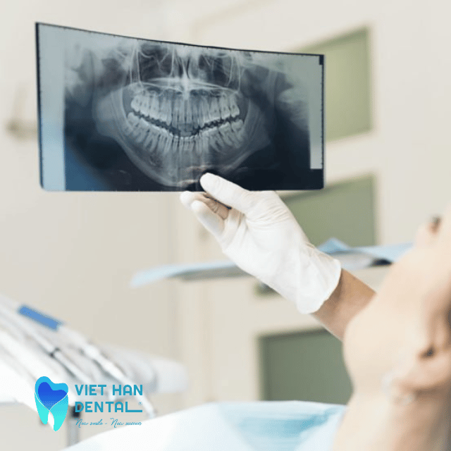
- Panoramic X-rays
- Cephalometric X-rays
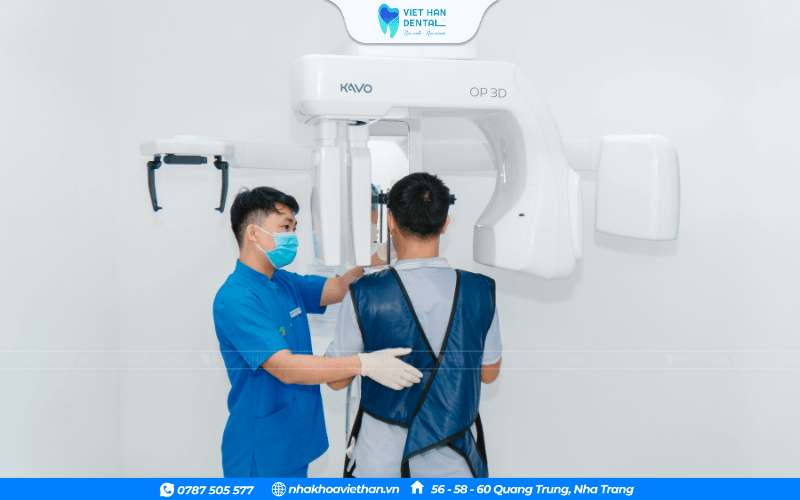
- 3D Cone Beam CT scans
- Digital intraoral scans
Not every patient requires every type of imaging. The dentist selects the appropriate method based on the clinical condition and treatment needs.
1. Detecting Problems That Are Difficult to See
A visual examination alone may not reveal every dental problem.
Dental X-rays can help identify conditions such as:
- Cavities between teeth
- Infection around tooth roots
- Impacted wisdom teeth
- Bone loss associated with gum disease
- Hidden dental structures
- Certain abnormalities of the jawbone
Early detection can help dentists recommend appropriate treatment before a problem becomes more advanced.
2. Supporting More Precise Treatment Planning
Dental imaging is especially important for treatments that require detailed assessment of the teeth, bone and surrounding anatomy.
For example, 3D Cone Beam CT may be used in implant planning to evaluate:
- Jawbone volume and anatomy
- Position of important anatomical structures
- Available bone for implant placement
- Relationship between the implant site and surrounding teeth
Digital scans can also support the planning of crowns, veneers, orthodontic treatment and other dental restorations.
3. Helping Patients Understand Their Dental Condition
Dental images can make complex information easier to understand.
Instead of relying only on verbal explanations, the dentist can show patients:
- Where a cavity is located
- Why a tooth may require treatment
- The condition of the surrounding bone
- How teeth are positioned
- What changes are expected during treatment
This allows patients to participate more confidently in decisions about their oral health.
4. Monitoring Treatment Progress
Images can also be useful for comparing oral conditions over time.
Depending on the treatment, dentists may use photographs, scans or X-rays to monitor:
- Orthodontic tooth movement
- Healing after dental surgery
- Changes in gum and bone condition
- Implant treatment progress
- The condition of existing restorations
These records help the dental team evaluate treatment and identify changes that may require attention.
5. Improving Communication Between Dental Professionals
Complex dental treatment often involves collaboration between dentists, specialists and dental laboratory technicians.
Digital images and scans can help communicate important clinical information, including tooth position, bite relationships and restoration design.
This is particularly useful for implant rehabilitation, orthodontics and aesthetic restorative dentistry.
Is Dental Imaging Safe?
Dental imaging should be performed only when clinically justified.
Modern digital X-ray systems are designed to use controlled radiation doses, while intraoral photographs and optical digital scans do not use ionising radiation.
Your dentist will consider your medical history, previous imaging and treatment needs before recommending an X-ray or CT scan.
Dental Imaging at Viet Han Dental Clinic
At Viet Han Dental Clinic, diagnostic technology supports our approach to careful examination and individualised treatment planning.
Depending on the clinical indication, our facilities include:
- 3D Cone Beam CT
- Digital panoramic and cephalometric imaging
- Digital intraoral scanning
- Intraoral photography
These technologies help our dentists assess oral conditions, communicate treatment options and plan procedures with greater detail.
Understanding Your Smile Starts With the Right Diagnosis
Dental images are valuable tools, but they do not replace a dentist’s clinical examination. The most reliable assessment combines your symptoms, medical and dental history, direct examination and appropriate diagnostic imaging.
If you are experiencing tooth pain, considering dental implants, planning orthodontic treatment or simply wish to understand your oral health more clearly, contact Viet Han Dental Clinic to arrange a consultation.
Viet Han Dental Clinic – Nha Trang
56–58–60 Quang Trung, Nha Trang, Vietnam
Hotline / WhatsApp: +84 787 505 577
Website: nhakhoaviethan.vn